
Diagnostic imaging occupies a significant place in gastroenterology. It provides a detailed look inside your body without surgery. As a powerful tool, it helps to pinpoint digestive issues accurately and efficiently. It includes various techniques – from the familiar X-rays to more sophisticated procedures like DEXA scan colorado. In this blog, we will delve into its importance and how it has revolutionized patient care in gastroenterology.
Understanding Diagnostic Imaging
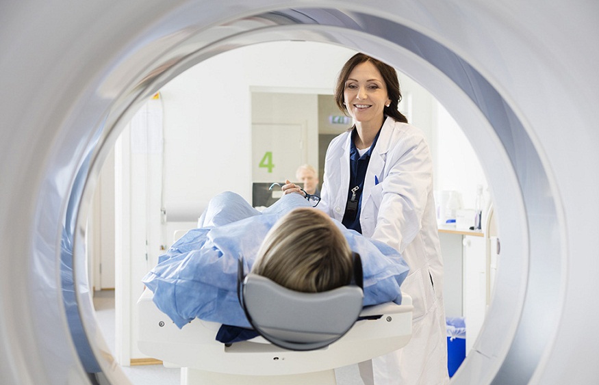
Imagine having a window into the body. That’s diagnostic imaging. It allows doctors to observe the body’s internal workings. The simplest form is the X-ray. It uses radiation to create images of bones and certain body tissues. More advanced forms include MRIs and CT scans. These use magnetic fields or radiation to create detailed images of soft tissues.
Connection to Gastroenterology
Gastroenterology deals with the digestive tract. This includes the stomach, intestines, and related organs. Diagnostic imaging can help doctors see these organs clearly. It can reveal problems like tumors, blockages, or inflammation. It can also guide treatments. For instance, in a procedure called an endoscopy, a doctor uses a tiny camera to see inside the body.
Types of Diagnostic Imaging in Gastroenterology
There are several kinds of diagnostic imaging used in gastroenterology. Each has its strengths.
- X-rays are good for seeing the shape and size of organs. They can reveal blockages or abnormal growths.
- CT scans provide more detail than X-rays. They can show the layers of the stomach wall or the small intestines.
- MRIs give the best images of soft tissues. They can show the thickness of the intestinal wall, useful in diagnosing conditions like Crohn’s disease.
- Ultrasounds can show the flow of blood through the vessels in the digestive tract. This is helpful in diagnosing conditions like liver disease.
- DEXA scans are primarily used to measure bone density. They can be used in gastroenterology to monitor patients with conditions that affect calcium absorption, like celiac disease.
Table: Comparison of Diagnostic Imaging Techniques
| Type | Strengths | Common Uses in Gastroenterology |
| X-ray | Shows shape and size of organs reveals blockages or abnormal growths | Diagnosis of tumors, ulcers |
| CT Scan | Shows more detail than X-rays, can view layers of organs | Diagnosis of appendicitis, pancreatitis |
| MRI | Gives detailed images of soft tissues | Diagnosis of Crohn’s disease, liver disease |
| Ultrasound | Shows blood flow through vessels | Diagnosis of gallstones, liver disease |
| DEXA Scan | Measures bone density | Monitoring of celiac disease |
Conclusion
In gastroenterology, diagnostic imaging plays a crucial role. It reveals what’s happening inside the body. It enables doctors to diagnose accurately, plan treatment, and monitor progress. It’s like a window into the body—a window that opens the way to better health.